


Bio-Imaging Technologies in Studying Osteoarticular Diseases
As a specialized Contract Research Organization, at AGINKO we have access to the latest technologies. We can cover a large range of bio-imaging techniques to evaluate bone resorption, formation, fracture and cartilage degeneration repair but also various other parameters.
We provide our clients with a translational pharmacological approach which gives quantifiable and visible evidence of disease progression and treatment, drug efficacy and safety in their animal models.
Xrays and Contact Radiography
X-rays technology is the oldest and most commonly used form of medical imaging and remains an important tool for the diagnosis of many osteoarticular disorders. We perform Standard Xrays as well contact Xrays (Faxitron).
 |
|
|
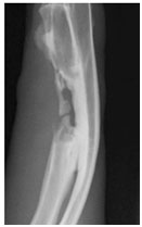
NZW rabbit ulnar critical size defect radiograph |
|
 |
|
|
 |
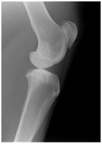
| Contact radiography of a Dog PLF model |
In Vivo Computed Tomography
Computed Tomography (CT) is an X-ray based technique imaging live animals. This technique is usually used to assess a surgical technique, to check the position of a medical device or to follow disease progression while a study is ongoing. At AGINKO we offer vivo CT for all species from small to large animals.
Micro-Computed Tomography
MicroComputed Tomography is an X-ray transmission imaging technique. It creates cross-sections of the physical samples that can be used to recreate a virtual model (3D model) without destroying the original object. This is useful for nondestructively visualizing and analyzing the internal structure of materials.
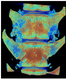
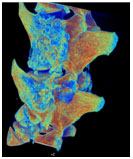
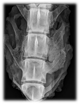
| 3D reconstruction of a fused dog spine |
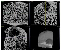
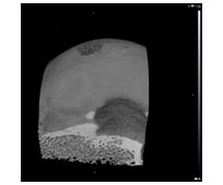
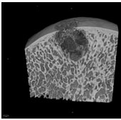
| 2D and 3D images of a condyle defect in a sheep knee. |
Magnetic Resonance Imaging (MRI)
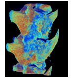
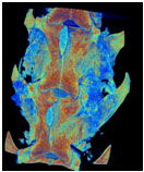
MRI is a medical imaging technique used to obtain pictures of the anatomy and the physiological processes of the body in both health and disease situation. At Aginko we use MRI to assess cartilage repair or disc degeneration both in vivo and ex vivo.
 |
|
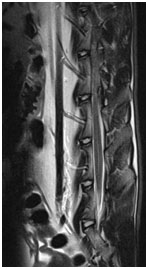
T2 sequence of a rabbit spine(3 Tesla) |
 |
|
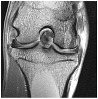
Radiograph of a goat kneedGEMRIC sequence of a goat knee (3 Tesla) |
In Vivo Bioluminescence Imaging
In vivo bioluminescent imaging (BLI) is a versatile and sensitive tool that is based on detection of light emission from cells. Optical imaging by bioluminescence allows a low-cost, noninvasive, and real-time analysis of disease processes in living organisms. Bioluminescence is used to track tumor cells, bacterial and viral infections, gene expression, and treatment response. At Aginko bioluminescence can be used to visualize the progression of bone cancer in spine or tibia of rodents.
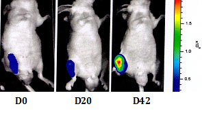
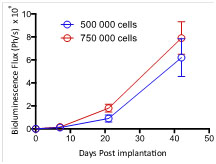
Prostate cancer cells were injected directly in the tibia of nude mice. Tumor burden was assessed by bioluminescence at D0, D20 and D42 after tumor cells injection.
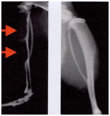
High resolution X-rays of ipsilateral (left) and contralateral (right) hindpaw of mice 35 days post injection of prostate cancer cells.